Cerebral edema, also known as brain swelling, is the
excess accumulation of fluid in intracellular or extracellular spaces of the
brain, causing an increase in pressure known as intracranial pressure.
Cerebral edema is one of the
most dangerous diseases that may affect humans. So we find many of us frightened
by the severity of the health complications that result from it, which may
radically affect the patient’s life.
So let us take a comprehensive look at
cerebral edema in terms of causes, symptoms, methods of diagnosis and
treatment.
Cerebral Edema (Brain Swelling) – Types, Symptoms, Causes, Diagnosis, and Treatment
 |
| Cerebral edema or brain swelling |
What is Cerebral Edema?
Cerebral edema,
also known as brain swelling, is a serious and life-threatening condition that
occurs as a result of fluid accumulating inside the brain, which increases the
size of the brain.
Because the brain is confined to the cranial box, so any
increase in the volume of fluid inside the brain raises intracranial pressure,
which reduces blood flow to the brain and weakens the supply of the brain with
oxygen.
Cerebral edema is sometimes difficult to treat and leads to permanent
damage.
also known as brain swelling, is a serious and life-threatening condition that
occurs as a result of fluid accumulating inside the brain, which increases the
size of the brain.
Because the brain is confined to the cranial box, so any
increase in the volume of fluid inside the brain raises intracranial pressure,
which reduces blood flow to the brain and weakens the supply of the brain with
oxygen.
Cerebral edema is sometimes difficult to treat and leads to permanent
damage.
Types of
Cerebral Edema
Vasogenic
edema: The most common type occurs as a result of a breakdown of the
blood-brain barrier due to trauma or external injury.
edema: The most common type occurs as a result of a breakdown of the
blood-brain barrier due to trauma or external injury.
Cytotoxic/ionic/cellular
edema: The blood-brain barrier remains healthy, but there is a
defect in the brain cells that makes it unable to produce energy properly.
edema: The blood-brain barrier remains healthy, but there is a
defect in the brain cells that makes it unable to produce energy properly.
Interstitial/hydrocephalic
edema: Interstitial/hydrocephalic edema is caused by hydrocephalus.
edema: Interstitial/hydrocephalic edema is caused by hydrocephalus.
Osmotic /
hypostatic edema: Osmotic edema occurs as a result of a loss of osmotic balance
between the blood plasma and the visceral tissue of the brain, which promotes
the accumulation of fluid within the brain.
hypostatic edema: Osmotic edema occurs as a result of a loss of osmotic balance
between the blood plasma and the visceral tissue of the brain, which promotes
the accumulation of fluid within the brain.
Hydrostatic
edema: Hydrostatic edema occurs as a result of a loss of the ability
to regulate blood circulation within the brain.
edema: Hydrostatic edema occurs as a result of a loss of the ability
to regulate blood circulation within the brain.
Cerebral Edema or Brain Swelling:
All you need to know
Brain swelling has many names:
- Brain edema
- Cerebral edema
- Elevated intracranial pressure
 |
| Increased intracranial pressure |
Increased Intracranial Pressure (ICP): an overview
Intracranial pressure (ICP) is the pressure that is formed in the component parts of the skull: the brain,
blood vessels and cerebrospinal fluid (CSF).
Intracranial pressure is equivalent to the pressure of
the cerebrospinal fluid that can be measured by a lumbar puncture.
The
normal CSF value is usually 8 mm Hg, or 110 mm h2o (80 – 180 mm h2o). This
value is variable and is related to several factors, such as body and head
position, heart and respiratory activity, effort, pressure in the veins and pain
stimuli.
blood vessels and cerebrospinal fluid (CSF).
Intracranial pressure is equivalent to the pressure of
the cerebrospinal fluid that can be measured by a lumbar puncture.
The
normal CSF value is usually 8 mm Hg, or 110 mm h2o (80 – 180 mm h2o). This
value is variable and is related to several factors, such as body and head
position, heart and respiratory activity, effort, pressure in the veins and pain
stimuli.
The skull
resembles a solid, closed box. Therefore, any increase in the size of any of
its components causes an increase in intracranial pressure.
resembles a solid, closed box. Therefore, any increase in the size of any of
its components causes an increase in intracranial pressure.
Elevated Intra –
Cranial Pressure (EICP)
Elevated Intra
– Cranial Pressure (EICP) or Intracranial hypertension, is a medical condition
that requires urgent treatment, usually with severe effects.
– Cranial Pressure (EICP) or Intracranial hypertension, is a medical condition
that requires urgent treatment, usually with severe effects.
High pressure
is not governed by a direct relationship with the change in volume.
The
increase in the size of one of the brain’s chambers, up to a certain extent,
does not cause an increase in pressure, due to the mechanisms of adjustment and
compensation found in the brain and the elasticity of the different tissues.
However, if this limit is exceeded, any slight increase in size causes a sharp
increase in intracranial pressure.
is not governed by a direct relationship with the change in volume.
The
increase in the size of one of the brain’s chambers, up to a certain extent,
does not cause an increase in pressure, due to the mechanisms of adjustment and
compensation found in the brain and the elasticity of the different tissues.
However, if this limit is exceeded, any slight increase in size causes a sharp
increase in intracranial pressure.
High
intracranial pressure may have dangerous consequences, due to a decrease in the
perfusion pressure in the brain, to the point where the blood supply to the
brain cells is interrupted.
intracranial pressure may have dangerous consequences, due to a decrease in the
perfusion pressure in the brain, to the point where the blood supply to the
brain cells is interrupted.
Low intracranial pressure: intracranial pressure may be lower than normal due to cerebrospinal fluid leakage as a result of a hole in the covers of the brain, spontaneously or as a result of shock (for example, after a lumbar puncture).
The most prominent symptom of this condition is a headache that gets worse when standing up.
Diagnosis is made if low pressure is detected in the lumbar puncture examination and then confirmed by magnetic resonance imaging (MRI) that shows (by dye) highlighting the brain envelopes with the indicated Gadolinium.
The most prominent symptom of this condition is a headache that gets worse when standing up.
Diagnosis is made if low pressure is detected in the lumbar puncture examination and then confirmed by magnetic resonance imaging (MRI) that shows (by dye) highlighting the brain envelopes with the indicated Gadolinium.
Cerebral perfusion pressure (CPP)
 |
| Cerebral Perfusion Pressure – an overview |
What is Cerebral
perfusion pressure (CPP)?
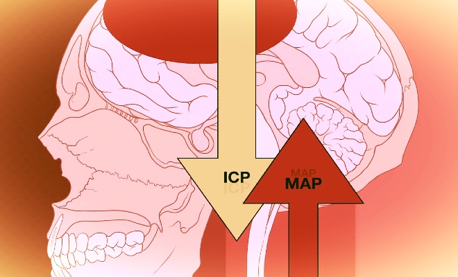
Cerebral
perfusion pressure (CPP) is the difference between the blood pressure in the arteries
of the brain (arterial pressure- MAP) and
the intracranial pressure (ICP) on these arteries, which determines the
transfer of oxygen and metabolites from the blood to the brain cells.
perfusion pressure (CPP) is the difference between the blood pressure in the arteries
of the brain (arterial pressure- MAP) and
the intracranial pressure (ICP) on these arteries, which determines the
transfer of oxygen and metabolites from the blood to the brain cells.
In a healthy
brain, there is a special mechanism that regulates the flow of blood
(Autoregulation) and maintains a continuous flow regardless of the level of
blood pressure, by changing the degree of resistance in the walls of blood
vessels.
This mechanism may be malfunctioned by the disorder caused by high
intracranial pressure, which makes the brain more sensitive to changes in blood
pressure.
brain, there is a special mechanism that regulates the flow of blood
(Autoregulation) and maintains a continuous flow regardless of the level of
blood pressure, by changing the degree of resistance in the walls of blood
vessels.
This mechanism may be malfunctioned by the disorder caused by high
intracranial pressure, which makes the brain more sensitive to changes in blood
pressure.
In contrast,
hypertension lowers the ischemic stroke, creating a life-threatening condition
for the brain.
Another dangerous consequence of high intracranial pressure
is the risk of herniation, i.e. pushing the content of brain tissue from
one of the compartments in the skull, where the pressure is high, to another
compartment where the pressure is low.
hypertension lowers the ischemic stroke, creating a life-threatening condition
for the brain.
Another dangerous consequence of high intracranial pressure
is the risk of herniation, i.e. pushing the content of brain tissue from
one of the compartments in the skull, where the pressure is high, to another
compartment where the pressure is low.
This is through narrow openings in
the covers of the brain or the base of the skull, along with increased pressure
and damage to vital areas of the brain.
The increase in
the size of any of the internal components of the skull, due to various
reasons, may cause intracranial hypertension:
the size of any of the internal components of the skull, due to various
reasons, may cause intracranial hypertension:
Cerebrospinal Fluid (CSF)
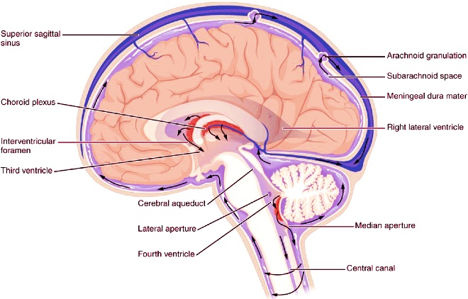
Cerebrospinal fluid is a clear, colorless liquid around your brain and spinal cord.
CSF is a
total volume of about 150 ml,
running in cavities inside (chambers) and around the brain and spinal cord (in
cisterna subarachnoidalis).
CSF helps protect the brain from shocks, in
regulating metabolic activity and removing exudate.
The fluid is absorbed in
the upper area of the brain surface into the vast sinuses, through microscopic
villi called Arachnoid villi.
CSF is a
total volume of about 150 ml,
running in cavities inside (chambers) and around the brain and spinal cord (in
cisterna subarachnoidalis).
CSF helps protect the brain from shocks, in
regulating metabolic activity and removing exudate.
The fluid is absorbed in
the upper area of the brain surface into the vast sinuses, through microscopic
villi called Arachnoid villi.
The delicate balance between fluid production, on the one hand, and its absorption, on the other hand, maintains its constant quantity.
The amount of fluid may increase
significantly, causing high pressure inside the skull, as a result of the
following:
The amount of fluid may increase
significantly, causing high pressure inside the skull, as a result of the
following:
1. Increased
CSF production – a very rare phenomenon, because the normal ability to absorb
CSF is greater than the ability to produce it.
CSF production – a very rare phenomenon, because the normal ability to absorb
CSF is greater than the ability to produce it.
2. Disorders of
cerebrospinal fluid (CSF) circulation – caused by bile duct obstructions as a result of birth defects,
infection, bleeding or increased growth, or any compressive growth elsewhere
nearby.
The fluid produced accumulates in the chambers above the site of
the blockage, causing them to swell and increase in size, which increases
intracranial pressure.
This condition is called obstructive hydrocephalus
and requires, in addition to removing the obstruction cause, treatment for
fluid drainage (Shunt), i.e. placing a tube from the side chamber, passing
through the subcutaneous canal and up to the abdominal cavity, to drain the
fluid.
cerebrospinal fluid (CSF) circulation – caused by bile duct obstructions as a result of birth defects,
infection, bleeding or increased growth, or any compressive growth elsewhere
nearby.
The fluid produced accumulates in the chambers above the site of
the blockage, causing them to swell and increase in size, which increases
intracranial pressure.
This condition is called obstructive hydrocephalus
and requires, in addition to removing the obstruction cause, treatment for
fluid drainage (Shunt), i.e. placing a tube from the side chamber, passing
through the subcutaneous canal and up to the abdominal cavity, to drain the
fluid.
3.
Cerebrospinal fluid absorption disorder caused by a blockage in Arachnoid Villi
due to inflammation, contamination or bleeding in the cerebrospinal fluid or
due to an unknown cause as a result of age, the formation of cerebral
edema is non-obstructive.
Cerebrospinal fluid absorption disorder caused by a blockage in Arachnoid Villi
due to inflammation, contamination or bleeding in the cerebrospinal fluid or
due to an unknown cause as a result of age, the formation of cerebral
edema is non-obstructive.
4. Increased
cerebral blood volume: Arterial capillaries in the brain are sensitive to
a change in the concentration of gases in the blood.
An increase in the
partial pressure of carbon dioxide, or a sharp decrease in the partial pressure
of oxygen due to breathing problems, will lead to its expansion and an increase
in the amount of blood (and pressure) in the brain.
A sharp rise in blood pressure
can disrupt the mechanisms of autoregulation and high pressure within the
skull, coinciding with the occurrence of hypertensive encephalopathy.
cerebral blood volume: Arterial capillaries in the brain are sensitive to
a change in the concentration of gases in the blood.
An increase in the
partial pressure of carbon dioxide, or a sharp decrease in the partial pressure
of oxygen due to breathing problems, will lead to its expansion and an increase
in the amount of blood (and pressure) in the brain.
A sharp rise in blood pressure
can disrupt the mechanisms of autoregulation and high pressure within the
skull, coinciding with the occurrence of hypertensive encephalopathy.
An increase in
the volume of venous blood in the brain can be caused by a blockage of the
drainage system from venous sinuses, as a result of thrombosis or a foreign
body airway obstruction (FBAO (tumor, fracture) or increased central venous
pressure transferred to the veins of the brain.
the volume of venous blood in the brain can be caused by a blockage of the
drainage system from venous sinuses, as a result of thrombosis or a foreign
body airway obstruction (FBAO (tumor, fracture) or increased central venous
pressure transferred to the veins of the brain.
5. Increase in
brain size: This phenomenon includes the main factors of intracranial
hypertension:
brain size: This phenomenon includes the main factors of intracranial
hypertension:
1. Operations
occupying space in the cranial cavity: tumor, sinus formation, bleeding, an
abscess. The pressure increases due to the increase in size without additional
space in the skull. This occurs due to
obstruction of the cerebrospinal fluid (CSF) leak or CSF absorption, due to
blockage of venous sinuses or the formation of cerebral edema.
occupying space in the cranial cavity: tumor, sinus formation, bleeding, an
abscess. The pressure increases due to the increase in size without additional
space in the skull. This occurs due to
obstruction of the cerebrospinal fluid (CSF) leak or CSF absorption, due to
blockage of venous sinuses or the formation of cerebral edema.
2. Brain tissue swelling due to fluid accumulation in it, due to a variety
of injuries: stroke, blockage, swelling, bleeding, infection, inflammation,
hypoxia, metabolic and toxic disorders, etc.
of injuries: stroke, blockage, swelling, bleeding, infection, inflammation,
hypoxia, metabolic and toxic disorders, etc.
 |
| Cerebrospinal fluid (CSF) function |
Symptoms of Cerebral Edema
Cerebral edema
is difficult to diagnose, but the presence of some symptoms and signs helps the
doctor in the diagnosis process, such as:
is difficult to diagnose, but the presence of some symptoms and signs helps the
doctor in the diagnosis process, such as:
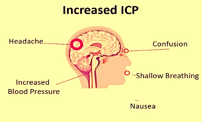
- Headache
- Dizziness
- Nausea
- Vomiting
- Numbness
- Loss of
muscular nervous coordination
In severe
cases, the patient may notice symptoms such as:
cases, the patient may notice symptoms such as:
- Mood changes
- Memory loss
- Vision loss
- Low heart rate
- High blood
pressure - Difficulty
speaking - Difficulty
moving - Neck pain
- Micturition
Reflex - Unconsciousness
- Epileptic
seizures
Symptoms of
cerebral edema vary depending on the severity of the swelling and the
underlying cause.
Dilated fundus examination (DFE) shows the presence of
papilledema. For children, the Fonticuli cranii are swollen and the cranial
sutures may separate.
cerebral edema vary depending on the severity of the swelling and the
underlying cause.
Dilated fundus examination (DFE) shows the presence of
papilledema. For children, the Fonticuli cranii are swollen and the cranial
sutures may separate.
Causes of Cerebral Edema
There are several reasons for cerebral edema such as:
Brain injuries: such as direct hits to the head may
cause damage to the brain. Severe injuries can lead to fractures of the skull
bones. The bone fragments resulting from the fractures may lead to a
rupture of the blood vessels, leading to cerebral edema.
cause damage to the brain. Severe injuries can lead to fractures of the skull
bones. The bone fragments resulting from the fractures may lead to a
rupture of the blood vessels, leading to cerebral edema.
Ischemic stroke: Some cases of
stroke lead to the occurrence of edema, especially if the cause of a
stroke is a decrease in the amount of oxygen delivered to the brain, which
leads to the death of brain cells and swelling as a result of the injury.
stroke lead to the occurrence of edema, especially if the cause of a
stroke is a decrease in the amount of oxygen delivered to the brain, which
leads to the death of brain cells and swelling as a result of the injury.
Brain hemorrhage: A brain
hemorrhage is a type of stroke that occurs when the blood vessels in the brain
rupture and leak. This leads to bleeding in or around the brain and causes
inflammation and an increase in intracranial pressure (ICP).
hemorrhage is a type of stroke that occurs when the blood vessels in the brain
rupture and leak. This leads to bleeding in or around the brain and causes
inflammation and an increase in intracranial pressure (ICP).
Infections: Some bacteria are able to cause
inflammation and swelling in the brain, especially if left untreated.
inflammation and swelling in the brain, especially if left untreated.
Brain tumors: the presence of tumors creates
additional pressure on the brain causing swelling.
additional pressure on the brain causing swelling.
Other causes of cerebral edema: Other
causes such as carbon monoxide (CO) poisoning, common hiking injuries, venomous
animal bites, viral infections, and some medications.
causes such as carbon monoxide (CO) poisoning, common hiking injuries, venomous
animal bites, viral infections, and some medications.
Diagnosis
of Cerebral Edema
Diagnosing cerebral edema is somewhat difficult and a challenge for
the attending physician, but by referring to symptoms and using some
techniques, a correct diagnosis can be made.
Some of the common procedures
that doctors use to diagnose cerebral edema include:
the attending physician, but by referring to symptoms and using some
techniques, a correct diagnosis can be made.
Some of the common procedures
that doctors use to diagnose cerebral edema include:
⇒Clinical examination of the patient to show the symptoms they
complain about.
complain about.
⇒Neurological examination to identify the extent and locate
idiopathic edema.
idiopathic edema.
⇒Computed tomography (CT) scan to locate the
swelling and identify the extent.
swelling and identify the extent.
⇒Magnetic resonance imaging (MRI) of the head to locate edema.
⇒Blood tests may help to determine the cause of cerebral edema.
Treatment
of Cerebral Edema
Cerebral edema is a life-threatening condition so you need to
monitor the patient’s condition in the intensive care room.
Early diagnosis and
treatment are necessary to save a patient’s life. Often the primary cause
must be treated quickly, using various medications to reduce the amount of
water in the brain and to produce cerebrospinal fluid.
The treatment aims
to restore blood and oxygen flow to the brain while reducing the swelling that
occurs at the same time.
The most common cerebral edema treatment options may
include:
monitor the patient’s condition in the intensive care room.
Early diagnosis and
treatment are necessary to save a patient’s life. Often the primary cause
must be treated quickly, using various medications to reduce the amount of
water in the brain and to produce cerebrospinal fluid.
The treatment aims
to restore blood and oxygen flow to the brain while reducing the swelling that
occurs at the same time.
The most common cerebral edema treatment options may
include:
Medications: Medications work to
reduce swelling and remove thrombus such as warfarin which thins the blood
and reduces the chances of blood clots. A thrombus is a blood
clot which closes the blood vessels and reduces blood flow and oxygen to
the brain.
reduce swelling and remove thrombus such as warfarin which thins the blood
and reduces the chances of blood clots. A thrombus is a blood
clot which closes the blood vessels and reduces blood flow and oxygen to
the brain.
Osmotherapy: Osmotherapy is a technique that pulls water out of the brain cells, which leads to the removal
of swelling and blood clots, stimulates blood circulation to the brain and
reduces intracranial pressure.
This is done by using osmotic solutions such
as mannitol and hypertonic saline.
of swelling and blood clots, stimulates blood circulation to the brain and
reduces intracranial pressure.
This is done by using osmotic solutions such
as mannitol and hypertonic saline.
Hyperventilation: Hyperventilation
aims to cause the patient to release more gas than he inhales and reduces the concentration of carbon dioxide in the bloodstream and reduces intracranial
pressure.
aims to cause the patient to release more gas than he inhales and reduces the concentration of carbon dioxide in the bloodstream and reduces intracranial
pressure.
Hypothermia: This type of treatment
involves lowering the body temperature, which can reduce the
metabolic activity of the brain and thus reduces swelling.
involves lowering the body temperature, which can reduce the
metabolic activity of the brain and thus reduces swelling.
Surgery: Surgery may include removing part of the skull and repairing
the damage, such as ruptured blood vessels. Severe cases of cerebral edema
(such as caused by tumors) may require surgery to reduce intracranial pressure.
the damage, such as ruptured blood vessels. Severe cases of cerebral edema
(such as caused by tumors) may require surgery to reduce intracranial pressure.
Ventriculostomy: The
ventriculostomy process aims to withdraw fluid from the brain by causing a hole
in the skull and inserting a tube to aspirate the fluid.
ventriculostomy process aims to withdraw fluid from the brain by causing a hole
in the skull and inserting a tube to aspirate the fluid.
Conclusion
Cerebral edema, or brain swelling, is a serious condition that requires prompt medical attention. It can result from various causes, including traumatic brain injury, stroke, infections, and tumors. The swelling increases pressure inside the skull, which can impair blood flow and oxygen delivery to the brain, leading to potential brain damage or even death if untreated.
There are several types of cerebral edema, including vasogenic, cellular, osmotic, and interstitial edema, each with distinct causes and mechanisms. Symptoms can range from headaches and nausea to seizures, visual disturbances, and changes in consciousness.
Treatment typically involves addressing the underlying cause and may include medications to reduce swelling, surgery to relieve pressure, and supportive care to manage symptoms. Early diagnosis and intervention are crucial for improving outcomes and preventing long-term complications.
Understanding the types, symptoms, causes, and treatment options for cerebral edema is essential for effective management and ensuring the best possible care for affected individuals.