Computed tomography (CT scan) is computer-aided medical imaging
using a series of X-ray waves to produce cross-sectional images of the body.
Computed tomography scan uses X-rays and a computer. A series of X-ray waves are passed
through the body at different angles to produce very detailed cross-sectional
images of the body called cross-sectional imaging.
CT scan gets detailed
information about organs without pain and can replace exploratory surgery in
many cases. Let’s learn more about computed tomography.
 |
| Computed Tomography (CT) scan is an X-ray diagnostic imaging technique that creates detailed pictures, or scans, of the body’s internal organs. |
Computed Tomography (CT) – A Diagnostic
Imaging Technique: How CT Scan works?
Imaging Technique: How CT Scan works?
What is a Computed Tomography (CT) Scan?
Computerized
Axial Tomography (CAT) or Computed Tomography (CT) is an X-ray
diagnostic imaging technique that forms a three-dimensional image of the
body’s internal organs from several two-dimensional images captured around a fixed axis of rotation.
This type of imaging is different from ordinary X-ray
imaging (Röntgen X-ray), which gives us a two-dimensional image, much
like the normal film. A full body scan is usually a type of computed tomography scan that scans your whole body.
Axial Tomography (CAT) or Computed Tomography (CT) is an X-ray
diagnostic imaging technique that forms a three-dimensional image of the
body’s internal organs from several two-dimensional images captured around a fixed axis of rotation.
This type of imaging is different from ordinary X-ray
imaging (Röntgen X-ray), which gives us a two-dimensional image, much
like the normal film. A full body scan is usually a type of computed tomography scan that scans your whole body.
CT
scan is
performed by a special device, called a CAT device or CT scanner that produces
multiple images.
CT scans of internal organs, bones, soft tissues, and blood
vessels provide more details than traditional X-rays and can be seen in several
horizontal or vertical sections complementary.
scan is
performed by a special device, called a CAT device or CT scanner that produces
multiple images.
CT scans of internal organs, bones, soft tissues, and blood
vessels provide more details than traditional X-rays and can be seen in several
horizontal or vertical sections complementary.
A CT scan radiation is performed in many cases to diagnose any change in the anatomical structure of the organs, whether at the level of the full organ or even tissue.
This rest
may diagnose damage to internal organs caused by various events such as trauma,
fractures, infections, tumors and other Space Occupying Lesions (SOL) that
cannot be seen with the naked eye or through simple visual imaging.
This rest
may diagnose damage to internal organs caused by various events such as trauma,
fractures, infections, tumors and other Space Occupying Lesions (SOL) that
cannot be seen with the naked eye or through simple visual imaging.
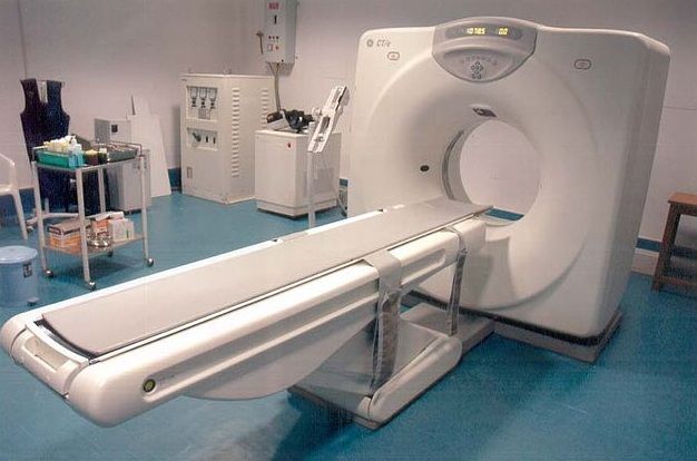
What is a CAT Scan Machine
Look Like?
According
to the National Institute of Biomedical Imaging and Bioengineering
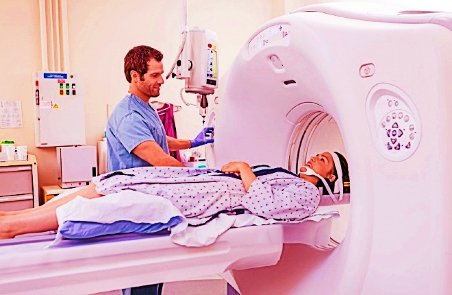
(NIBIB), During a Computerized Tomography – CT scan, the patient lays
down on the scanner bed – a square-shaped device in its vacuum center that
moves through the gantry – a doughnut-like ring that has an X-ray tube.
to the National Institute of Biomedical Imaging and Bioengineering
(NIBIB), During a Computerized Tomography – CT scan, the patient lays
down on the scanner bed – a square-shaped device in its vacuum center that
moves through the gantry – a doughnut-like ring that has an X-ray tube.
The
bed moves during the shooting process, both inside and outside the device.
The
patient is also asked not to move during the examination, and the imaging
technician gives the patient specific instructions on how to proceed during the
examination.
bed moves during the shooting process, both inside and outside the device.
The
patient is also asked not to move during the examination, and the imaging
technician gives the patient specific instructions on how to proceed during the
examination.
A shooting process is the release of narrow beams of X-rays across the
body.
X-rays are directly captured by digital detectors in contrast
to the source.
body.
X-rays are directly captured by digital detectors in contrast
to the source.
When
the X-ray source completes a full cycle, a sophisticated computer makes a
two-dimensional image of that segment of the body, which usually ranges from 0.04
to 0.4 inches thick.
The computer then combines and adds several
two-dimensional slices to create a three-dimensional image of the body,, which
makes it easy for the doctor to locate the patient’s problem.
the X-ray source completes a full cycle, a sophisticated computer makes a
two-dimensional image of that segment of the body, which usually ranges from 0.04
to 0.4 inches thick.
The computer then combines and adds several
two-dimensional slices to create a three-dimensional image of the body,, which
makes it easy for the doctor to locate the patient’s problem.
The scan usually takes less than 15 minutes depending on the area of the body that
has been imaged. In order to make it easier to identify any physical
abnormalities, the contrast material can be given to the patient.
Solutions that contain contrast materials such as barium or iodine are
introduced into the body, either rectally, orally or directly injected into the
bloodstream, on the basis of the target tissue.
The materials available
in the solution perform tasks temporarily by altering how X-rays interact with
some tissues of the body, causing the tissues to look different in the
resultant image.
The contrast offers basic ideas to the doctor and helps
him differentiate between normal and abnormal tissue.
has been imaged. In order to make it easier to identify any physical
abnormalities, the contrast material can be given to the patient.
Solutions that contain contrast materials such as barium or iodine are
introduced into the body, either rectally, orally or directly injected into the
bloodstream, on the basis of the target tissue.
The materials available
in the solution perform tasks temporarily by altering how X-rays interact with
some tissues of the body, causing the tissues to look different in the
resultant image.
The contrast offers basic ideas to the doctor and helps
him differentiate between normal and abnormal tissue.
How Does CT Scan Machine Work?
Computerized
Axial Tomography (CAT) or simply Computed Tomography (CT) is a modern medical
device that uses X-rays to obtain a stereoscopic image of the human body
instead of conventional X-rays that provide simple information about the skeletal structure of humans and some other organisms.
Axial Tomography (CAT) or simply Computed Tomography (CT) is a modern medical
device that uses X-rays to obtain a stereoscopic image of the human body
instead of conventional X-rays that provide simple information about the skeletal structure of humans and some other organisms.
CT
scanner is
a scanning device that produces X-rays.
X-rays are high-energy rays that
penetrate the living tissues of the human body and do not penetrate the bones.
X-rays are part of the electromagnetic spectrum, which consists of
light-emitting photons of 300,000 kilometers per second with high frequency and
far greater energy than visible light.
scanner is
a scanning device that produces X-rays.
X-rays are high-energy rays that
penetrate the living tissues of the human body and do not penetrate the bones.
X-rays are part of the electromagnetic spectrum, which consists of
light-emitting photons of 300,000 kilometers per second with high frequency and
far greater energy than visible light.
In
normal x-ray imaging, the X-ray machine works on a specific part of the human
body and the radiation from the body is received on the opposite side of a
special film.
The image is taken from the shadow of this radiation on the human
body and as it penetrates the living tissues of the body and does not penetrate
the bones and does not work on soft tissues.
The shadow is a picture of the
bone, and since the shadow is a picture in two dimensions, it does not give a complete idea of the shape of the body.
normal x-ray imaging, the X-ray machine works on a specific part of the human
body and the radiation from the body is received on the opposite side of a
special film.
The image is taken from the shadow of this radiation on the human
body and as it penetrates the living tissues of the body and does not penetrate
the bones and does not work on soft tissues.
The shadow is a picture of the
bone, and since the shadow is a picture in two dimensions, it does not give a complete idea of the shape of the body.
CT scan is a developed form of X-ray imaging and its development depends on the rapid development of the computer and its speed.
Using CAT devices, the doctor
can examine and diagnose the human body with precision so that it can look at
the human body as a component of thin slices to determine the disease and place it accurately and at high speed.
Using CAT devices, the doctor
can examine and diagnose the human body with precision so that it can look at
the human body as a component of thin slices to determine the disease and place it accurately and at high speed.
 |
| A computed tomography scan allows doctors to see inside the body. |
What are the Uses of Computed Tomography?
⇒CT scan is the fastest and most
accurate tool for examining the chest, abdomen, and pelvic imaging and provides
detailed images and transverse sections of all tissue types.
accurate tool for examining the chest, abdomen, and pelvic imaging and provides
detailed images and transverse sections of all tissue types.
⇒CT scan is used to examine patients with
damage or injuries when accidents such as vehicle accidents occur.
damage or injuries when accidents such as vehicle accidents occur.
⇒CT scan helps to determine the cause of
pain and difficulty in patients suffering from severe chest or abdominal
pain – stomachache or difficulty in breathing.
pain and difficulty in patients suffering from severe chest or abdominal
pain – stomachache or difficulty in breathing.
⇒CT scan is used to detect cancer in the
chest, abdomen, and pelvis, such as lymphoma, lung cancer, etc. because it
allows the doctor to confirm the presence of the tumor, measure its size, find
its exact location and determine its spread in other adjacent tissues.
chest, abdomen, and pelvis, such as lymphoma, lung cancer, etc. because it
allows the doctor to confirm the presence of the tumor, measure its size, find
its exact location and determine its spread in other adjacent tissues.
⇒CT scan helps to discover, diagnose and
treat vascular diseases that can lead to stroke, kidney failure, or even death.
treat vascular diseases that can lead to stroke, kidney failure, or even death.
⇒CT scan helps to assess pulmonary
embolism (blood clots in the lung vessels) as well as to examine aneurysms in
the aorta.
embolism (blood clots in the lung vessels) as well as to examine aneurysms in
the aorta.
⇒CT scan is used in the tomography of the
brain and head to evaluate the different structures of the brain to look for
brain tumors, stroke, bleeding areas, or vascular diseases. It is also
sometimes used to look at the skull.
brain and head to evaluate the different structures of the brain to look for
brain tumors, stroke, bleeding areas, or vascular diseases. It is also
sometimes used to look at the skull.
⇒CT scan is used in neck imaging to
examine the soft tissue in the neck and study the mass in the neck or to look
for enlarged lymph nodes or other glands.
examine the soft tissue in the neck and study the mass in the neck or to look
for enlarged lymph nodes or other glands.
⇒Computed tomography of the sinuses
is performed to diagnose sinus disease and to detect narrowing or obstruction
in the sinus drainage pathway.
is performed to diagnose sinus disease and to detect narrowing or obstruction
in the sinus drainage pathway.
⇒Spinal imaging is commonly used to
detect cartilage slide or narrowing of the spinal canal (narrowing of the
spine) in people with neck, arm, back and/or leg pain. It is also used to
detect the fracture of the spine.
detect cartilage slide or narrowing of the spinal canal (narrowing of the
spine) in people with neck, arm, back and/or leg pain. It is also used to
detect the fracture of the spine.
Computed tomography (CT) for
children – Paediatric CT scan
In
children, a CT scan is used to assess the following:
children, a CT scan is used to assess the following:
- Cancer of the lymph nodes.
- Kidney tumors.
- Congenital malformations of the
heart, kidneys and blood vessels. - Cystic fibrosis.
- Complications of acute
appendicitis. - Complications of pneumonia.
- Inflammatory bowel disease.
- Serious injuries (such as
injuries caused by traffic accidents or sliding).
Procedures before computed tomography (CT) scan
⇨You should wear comfortable and
loose clothing you may be asked to wear a hospital gown.
loose clothing you may be asked to wear a hospital gown.
⇨Remove any jewelry and clothing
that contains metal parts, glasses, and hairpins.
that contains metal parts, glasses, and hairpins.
⇨If the test needs to use a contrast
material, you should refrain from eating and drinking before the test for
several hours.
material, you should refrain from eating and drinking before the test for
several hours.
⇨Women should tell their doctor if
they are pregnant or have a potential for pregnancy.
they are pregnant or have a potential for pregnancy.
⇨Tell your doctor about any recent
diseases or other medical conditions and whether you have a history of heart
disease, asthma, diabetes, kidney disease or thyroid problems, because
the use of a contrast agent in these cases may be severe if these conditions
are not observed.
diseases or other medical conditions and whether you have a history of heart
disease, asthma, diabetes, kidney disease or thyroid problems, because
the use of a contrast agent in these cases may be severe if these conditions
are not observed.
⇨Before performing most CT scans of
the abdomen and pelvis, it is important to drink the oral contrast material
that contains the diluted barium.
This contrast material helps the radiologist
see the gastrointestinal tract (stomach, small intestine, and large intestine),
detect abnormalities in these organs, and separate these structures from other
structures within the abdomen.
the abdomen and pelvis, it is important to drink the oral contrast material
that contains the diluted barium.
This contrast material helps the radiologist
see the gastrointestinal tract (stomach, small intestine, and large intestine),
detect abnormalities in these organs, and separate these structures from other
structures within the abdomen.
Category at risk
Pregnant
women are not recommended for this test since the radiation from the scanner
maybe dangerous to the fetus. Consequently, this screening is not permitted
for pregnant women except in cases where other possibilities are not available.
There is no reason to do this for patients using a pacemaker or defibrillator.
However, patients with kidney failure must inform their doctor about their
illness prior to this test, because the Contrast may be dangerous to their
lives and cause further damage to the kidneys.
Patients who have previously
suffered from allergies to allergens or who are allergic to iodine should
inform the doctor.
Patients with diabetes, heart disease or thyroid disease
should also let know the doctor because they are more likely to have allergic
reactions to iodine.
women are not recommended for this test since the radiation from the scanner
maybe dangerous to the fetus. Consequently, this screening is not permitted
for pregnant women except in cases where other possibilities are not available.
There is no reason to do this for patients using a pacemaker or defibrillator.
However, patients with kidney failure must inform their doctor about their
illness prior to this test, because the Contrast may be dangerous to their
lives and cause further damage to the kidneys.
Patients who have previously
suffered from allergies to allergens or who are allergic to iodine should
inform the doctor.
Patients with diabetes, heart disease or thyroid disease
should also let know the doctor because they are more likely to have allergic
reactions to iodine.
Related diseases:
There
are many medical conditions, ranging from emergency medical conditions
resulting from accidents and shocks, loss of consciousness, internal injury
(ruptures/bleeding), chronic diseases, infections, infectious diseases, blood
clots, tumors, metastases, and much other damage to tissues.
are many medical conditions, ranging from emergency medical conditions
resulting from accidents and shocks, loss of consciousness, internal injury
(ruptures/bleeding), chronic diseases, infections, infectious diseases, blood
clots, tumors, metastases, and much other damage to tissues.
After Your Computed Tomography
(CT) Scan
In
general, there is no need for special procedures after the examination. The
only risks associated with this type of examination may be short-term exposure
to the radiation and/or allergic reactions to the substances (mostly due to
iodine sensitivity).
general, there is no need for special procedures after the examination. The
only risks associated with this type of examination may be short-term exposure
to the radiation and/or allergic reactions to the substances (mostly due to
iodine sensitivity).
An allergic reaction can be a rash, swelling in the neck area, or in some severe
cases of shortness of breath. Patients who notice these symptoms should
immediately notify their doctor.
Typically, the patient is required to wait for
up to half an hour after the examination, in order to ensure that he does not
suffer from immediate side effects.
cases of shortness of breath. Patients who notice these symptoms should
immediately notify their doctor.
Typically, the patient is required to wait for
up to half an hour after the examination, in order to ensure that he does not
suffer from immediate side effects.
Analysis of the Results
The
results of the CT scan are obtained after a computerized analysis and by a
radiologist.
Analysis of the results of the test requires some time, usually
with 130 images, and in some cases, 1000 images can be obtained.
The results
indicate the anatomy of the anatomical structure or the problems observed if
any, including Bleeding, Space-occupying Lesions (SOL) and others.
results of the CT scan are obtained after a computerized analysis and by a
radiologist.
Analysis of the results of the test requires some time, usually
with 130 images, and in some cases, 1000 images can be obtained.
The results
indicate the anatomy of the anatomical structure or the problems observed if
any, including Bleeding, Space-occupying Lesions (SOL) and others.